
A team in the Department of Biomedical Engineering at Texas A&M University, co-led by associate professor Akhilesh Gaharwar and assistant professor Abhishek Jain, has designed a 3D-bioprinted model of a blood vessel that can mimic its state of health and disease. This model could help in the development of novel cardiovascular drugs.
Vascular diseases such as aneurysms, peripheral artery disease and clots inside blood vessels account for 31% of global deaths. Despite this clinical burden, cardiovascular drug advances have slowed over the past 20 years. This decrease in cardiovascular therapeutic development is attributed to the lack of efficiency in converting possible treatments into approved methods, specifically due to the discrepancy between studies that take place outside the body compared to inside.
The team's research at Texas A&M University aims to remodel current methodologies to minimize this gap and improve the translatability of these techniques by applying 3D bioprinting to vascular medicine. Gaharwar is a biomaterials expert who has developed novel bioinks that offer unprecedented biocompatibility and control of mechanical properties needed for printing blood vessels. Jain’s expertise lies in creating biomimetic in vitro models of vascular and hematological diseases. The team reports its work in a paper in Advanced Healthcare Materials.
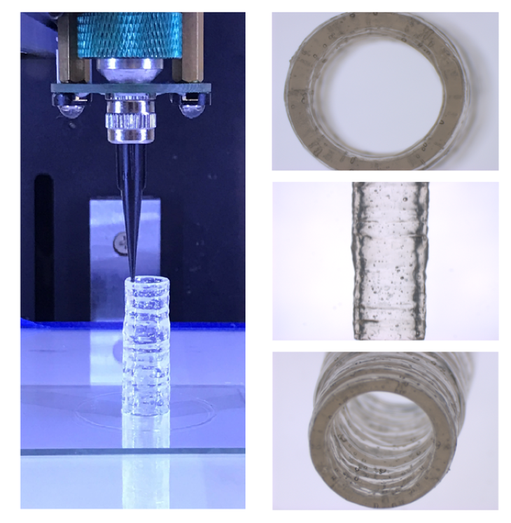
Bioprinting in 3D is an advanced manufacturing technique capable of producing unique, tissue-shaped constructs in a layer-by-layer fashion with embedded cells, making the arrangement more likely to mirror the native, multicellular makeup of vascular structures. A range of hydrogel bioinks have been developed for printing these structures, but there is a lack of bioinks that can mimic the vascular composition of native tissues. Current bioinks lack high printability and are unable to deposit a high density of living cells into complex 3D architectures.
To overcome these shortcomings, Gaharwar and Jain developed a new nanoengineered hydrogel-based bioink to print 3D, anatomically accurate, multicellular blood vessels. Their approach offers improved real-time resolution of both the macro-structure and tissue-level micro-structure, something that is not possible with current bioinks.
“A remarkably unique characteristic of this nanoengineered bioink is that regardless of cell density, it demonstrates a high printability and ability to protect encapsulated cells against high shear forces in the bioprinting process,” Gaharwar said. “Remarkably, 3D-bioprinted cells maintain a healthy phenotype and remain viable for nearly one month post-fabrication.”
Leveraging these unique properties, the nanoengineered bioink can be used to print 3D cylindrical blood vessels, consisting of living co-cultures of endothelial cells and vascular smooth muscle cells, which provides researchers the opportunity to model vascular function and disease impact. This 3D-bioprinted vessel provides a potential tool to understand vascular disease pathophysiology and assess therapeutics, toxins or other chemicals in preclinical trials.
This story is adapted from material from Texas A&M University, with editorial changes made by Materials Today. The views expressed in this article do not necessarily represent those of Elsevier. Link to original source.


