
A team led by bioengineers at the University of California, Los Angeles (UCLA) has developed a technique that uses a specially adapted 3D printer to build therapeutic biomaterials from multiple materials. This advance could represent a step toward on-demand printing of complex artificial tissues for use in transplants and other surgeries.
“Tissues are wonderfully complex structures, so to engineer artificial versions of them that function properly, we have to recreate their complexity,” said Ali Khademhosseini, who led the study and is a professor of engineering at the UCLA Samueli School of Engineering. “Our new approach offers a way to build complex biocompatible structures made from different materials.” The study is reported in a paper in Advanced Materials.
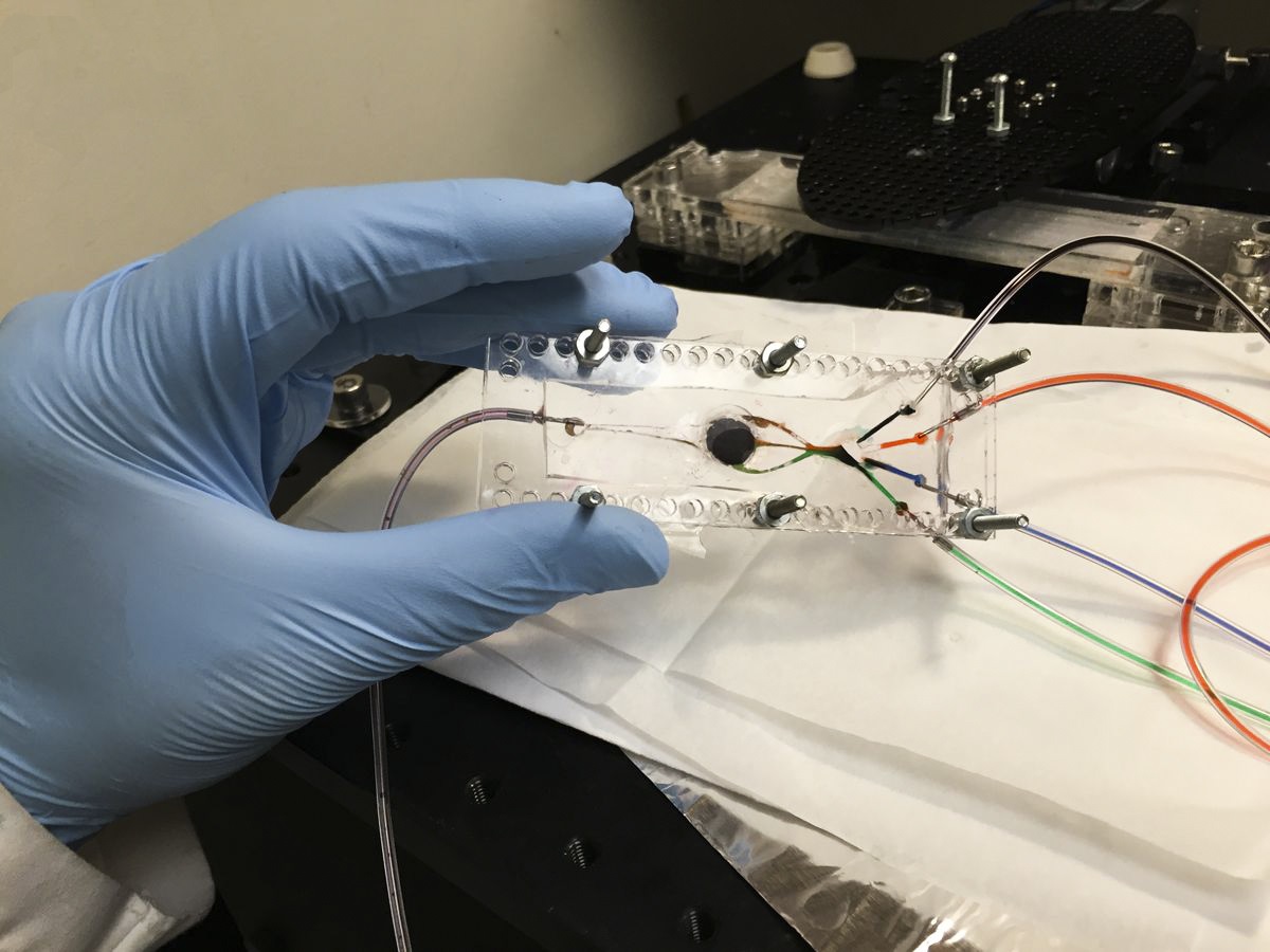
The novel technique uses a light-based printing process called stereolithography, and takes advantage of a customized 3D printer designed by Khademhosseini that has two key components. The first is a custom-built microfluidic chip – a small, flat platform similar in size to a computer chip – with multiple inlets that each ‘prints’ a different material. The other component is a digital micromirror, an array of more than a million tiny mirrors that each moves independently.
The researchers used different types of hydrogels – materials that, after passing through the printer, form scaffolds for tissue to grow into. The micromirrors direct light onto the printing surface, with the illuminated areas defining the outline of the 3D object that’s being printed. The light also triggers molecular bonds to form in the hydrogels, causing them to firm into solid materials. As the 3D object is printed, the mirror array changes the light pattern to indicate the shape of each new layer.
This process is the first to use multiple materials for automated stereolithographic bioprinting – an advance over conventional stereolithographic bioprinting, which only uses one type of material. While the demonstration device used four types of bio-inks, the study’s authors say that the process could accommodate as many inks as needed.
The researchers first used the process to make simple shapes, such as pyramids, before moving on to complex 3D structures that mimicked parts of muscle tissue and muscle-skeleton connective tissues. They also printed shapes mimicking tumors with networks of blood vessels, which could be used as biological models to study cancers. They tested these printed structures by implanting them in rats; the structures were not rejected.
This story is adapted from material from UCLA, with editorial changes made by Materials Today. The views expressed in this article do not necessarily represent those of Elsevier. Link to original source.

